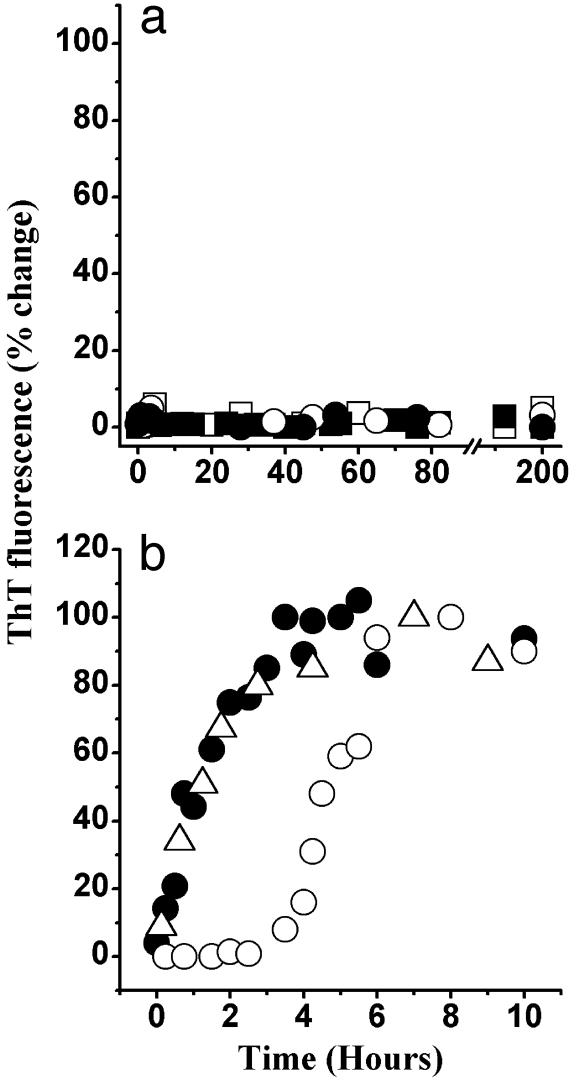
Fig. 6.
Time course of fibrillization of huPrP N-terminal fragments monitored by ThT fluorescence. (a) huPrP23–124 (squares) and huPrP23–137 (circles) incubated in the absence (open symbols) or presence (filled symbols) of huPrP23–144 seeds. (b) huPrP23–141 in the absence of preformed seeds (○) and in the presence of huPrP23–141 seeds (•) or huPrP23–144 seeds (▵). All samples were incubated at a concentration of 400 μM under identical conditions to those described in legend to Fig. 1. In case of seeded reactions, the amount of preformed fibrillar seeds was 1% wt/wt.

