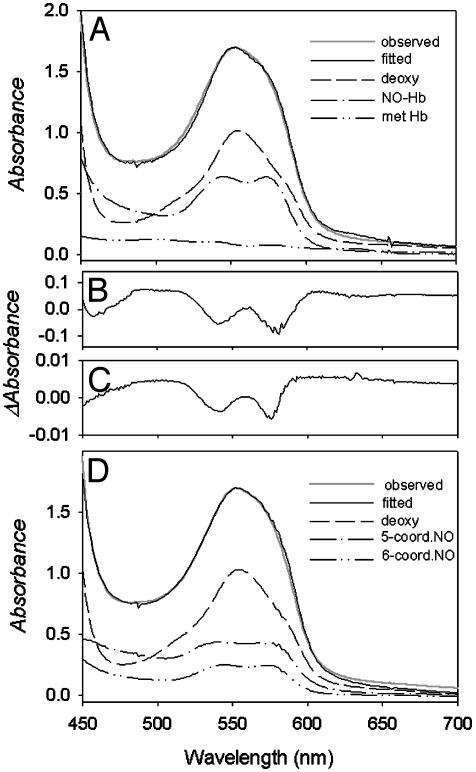
Fig. 2.
Analysis of visible absorbance spectra for samples at low NO to Hb ratios. (A) Comparison of observed and fitted spectra of partially nitrosylated 0.7 mM Hb in 0.05 M Bis-Tris/0.5 mM EDTA, and in the presence of the MetHb reductase system and DPG by using set I spectral standards as described in Materials and Methods. The relative contribution of each standard spectrum is indicated. (B) The large residual spectrum observed for similar samples when the MetHb standard was omitted from set I standards. In this case the samples contained dithionite in addition to MetHb reductase, precluding the presence of authentic MetHb. The magnitude of this residual spectrum depends on anions (IHP > DPG > Bis-Tris) and pH (low pH > high pH). The spectrum's shape is identical to that resulting from addition of IHP to a solution of hexacoordinate NO–Hb (4). (C) The residual spectrum for a Hb sample at pH 7.5 containing oxy Hb and MetHb that was fitted by using only the oxy Hb standard. (D) Comparison of observed and fitted spectra of partially nitrosylated Hb under conditions described in A by using set II spectral standards as described in Materials and Methods.

