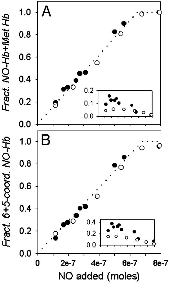
Fig. 3.
Stoichiometric NO binding at various NO to Hb ratios. Samples of 0.7 mM deoxy Hb in 0.05 M Bis-Tris/0.5 mM EDTA, and the MetHb reductase system, in the absence (○) and presence (•) of DPG at pH 7.5, 20°C, were equilibrated with varied amounts of NO gas. The NO fractional saturation was calculated from the visible absorbance spectra analyzed with set I (A) or set II (B) spectral standards. The dotted line indicates the level of NO–Hb expected for stoichiometric binding of NO to deoxy Hb. Stoichiometric binding of NO is evident when the fractional NO saturation is calculated from the sum of contributions from the NO–Hb standard spectrum and the MetHb standard (A), and from the sum of contributions from hexacoordinate and pentacoordinate NO–Hb (B). Insets show the apparent MetHb fractional saturation (A) and the fractional saturation of pentacoordinate NO (B).

