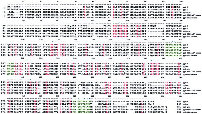
Figure 4.
Multiple sequence alignment of the PGI superfamily. Amino acid sequences of Bacillus PGI-B, Bacillus PGI-A, and PGI from pig, human, and mouse were aligned. Residues that are identical among all five sequences are highlighted in red. Positions of deletions are indicated by dashes. Two signature patterns ([LIVM]-G-G-R-[FY]-S-[LIVM]-x-[ST]-A-[LIVM]-G and [FY]-D-Q-x-G-V-E-x-x-K) of the PGI superfamily are colored in green. Amino acids that form the putative substrate-binding sites are denoted by asterisks.

