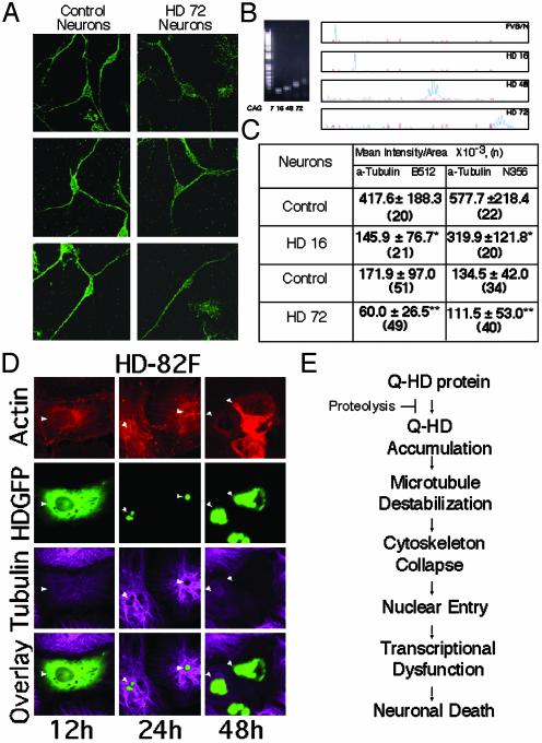
Fig. 6.
TB staining is diminished in cells expressing mhtt. (A) α-TB staining detected by using multiple C-terminal antibodies is diminished in neurons from HD72 transgenic mice. (B) Verification of repeat lengths in control (FVB/N) and transgenic mice expressing human htt with 16, 48, and 72 glutamines. CAG repeat length was determined by mobility on agarose gels (Left) and genescan analysis (Right). (C) Quantification of α-TB staining (by antibodies from A) in STR from control (FVB/N) and HD mice. Values are mean ± SD. *, P < 0.001; **, P < 0.01. (n) is number of cells observed. Experiment was repeated five times. (D) Microtubule structure is disrupted with time in CV-1 cells transfected with HD-82F. (E) Proposed mechanism for toxicity in HD.

