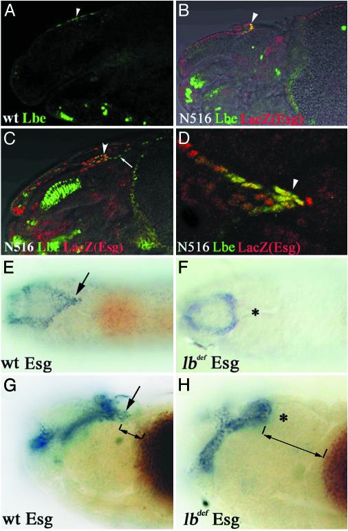
Fig. 2.
HANCs originate from the head epidermis and are not specified in lb-deficient embryos. (A–D) Confocal micrographs combined with transmitted light channels showing lateral (A–C) and dorsal (D) views of the head region of WT (A) and escargot enhancer trap N516 (B–D) embryos double-labeled for Lb (green) and β-galactosidase (red). (A) In the stage 11 embryo, a few cells in the dorsal head epidermis express lb (arrowhead). At stage 14 (B) and stage 16 (C), the lb-positive cells coexpress escargot (arrowheads) and localize to the tip of the invaginating frontal sac. (D) Dorsal view of stage 16 N516 embryo, showing the coexpression of lb and LacZ in the distal part of the frontal sac (arrowhead). (E and F) Nomarski micrographs representing the dorsal views of stage 16 embryos and (G and H) dorsolateral views of stage 14 embryos stained for escargot transcripts. (E and G) esg expression in WT embryos. The arrow indicates the HANCs. (F and H) In lb-deficient embryos, esg expression in the tip of the frontal sac is absent (*). The distance between the most distal frontal sac cells and the yolk sac (displaying brown background color) is much larger in lb-deficient embryos (compare bidirectional arrows in G and H). (Magnification: A and B, ×300; C, ×350; D, ×600; E and F, ×100; G and H, ×350.)

