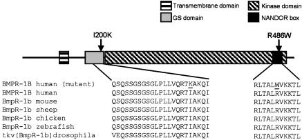
Fig. 3.
Schematic BMPR1B structure and mutations. Shown is the structure of BMPR1B showing the functional domains and the position of mutations (indicated by arrows). The amino acid sequences of BMPR1B are from different species, demonstrating the high degree of conservation within both domains. The two different mutations within the domains are underlined.

