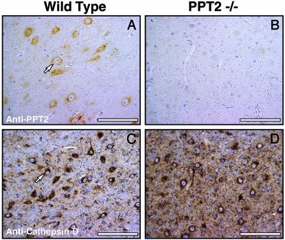
Fig. 1.
PPT2 immunoreactivity in WT and PPT2 knockout mouse brain. WT (A and C) and PPT2 knockout (B and D) mouse brain tissue was incubated with Abs directed against rat PPT2 (A and B) or cathepsin D (C and D). The punctate perinuclear staining pattern in cell bodies of neurons stained with anti-PPT2 Abs is similar to that of the lysosomal marker, cathepsin D. (Bar = 100 μm).

