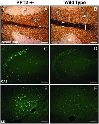
Fig. 3.
Neuropathology in PPT2 knockout mice. Light microscopic overview of silver-stained cerebellar folium in PPT2 knockout (A) and WT (B) mice. Atrophy of the granule cell layer and white tracts (white bars) and loss of dendritic arborization in the granule cell layer in the PPT2 knockout mice are shown. gcl, granular cell layer; ml, molecular layer. Arrows denote Purkinje neurons, which are well preserved. (C–F) Autofluorescent images of brain regions of 15-mo-old PPT2 knockout and WT mice. Regions shown are CA2 region of hippocampus (C and D) and lateral dorsothalamic (LD) nucleus (E and F). (Bar = 100 μm.)

