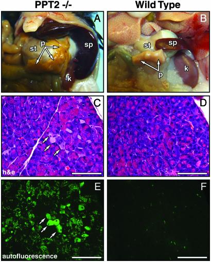
Fig. 4.
Visceral pathology in PPT2 knockout mice. (A and B) Viscera of PPT2 knockout and WT mice are shown in situ. Note the orange discoloration of the pancreas and the massive splenic enlargement in the knockout mouse. (C and D) Hematoxylin/eosin-stained sections of pancreas. Interstitial macrophages are increased (arrows) and cytoplasmic inclusions are seen intermixed with zymogen granules in exocrine cells. (E and F) Corresponding autofluorescent images. st, stomach; p, pancreas; sp, spleen; k, kidney. (Bar = 100 μm.)

