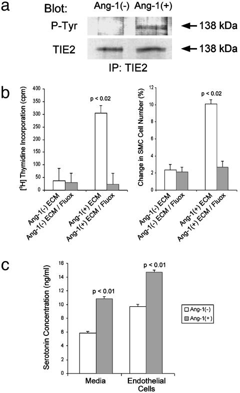
Fig. 4.
Ang-1-treated human pulmonary endothelial cells release a potent smooth muscle cell growth factor: serotonin. (a) Immunoblot analysis of anti-TIE2 immunoprecipitates from subcultured pulmonary arteriolar endothelial cells either treated or not treated with Ang-1 protein. Cells incubated with Ang-1 protein demonstrated TIE2 receptor phosphorylation, whereas untreated cells did not (Upper). Protein levels of TIE2 were similar in the two groups (Lower). (Upper) Immunoprecipitated (IP) with anti-TIE2 antibody, blotted with anti-phosphotyrosine (P-Tyr) antibody. (Lower) IP with anti-TIE2, blotted with anti-TIE2. (b) Stimulation of pulmonary smooth muscle cell proliferation by serum-free medium from Ang-1-treated endothelial cells is blocked by a serotonin transporter inhibitor. (Left)[3H]Thymidine incorporation in smooth muscle cells treated with (i) medium from endothelial cells not treated with Ang-1 [Ang-1(–)ECM], (ii) medium from endothelial cells not treated with Ang-1 to which 1 μM fluoxetine was added [Ang-1(–)ECM/Fluox], (iii) medium from endothelial cells treated with Ang-1 [Ang-1(+)ECM], or (iv) medium from endothelial cells treated with Ang-1 to which 1 μM fluoxetine was added [Ang-1(+)ECM/Fluox]. (Right) Percent change in smooth muscle cell count after treatment with the same media combinations as in Left.(c) Pulmonary endothelial cells treated with 50 ng/ml Ang-1 produce and secrete serotonin. Graph depicts results of ELISA quantitation of serotonin from cells treated with or without Ang-1 (right bars) and from media taken from cells treated with or without Ang-1 (left bars).

