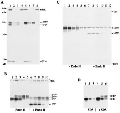
Figure 1.
Biosynthesis, assembly, and intracellular transport of HFE and TfR. (A) Association of β2m and TfR with HFE. LS10 cells grown for 48 h in the absence of tetracycline were labeled with [35S]methionine for 1 h (lanes 1–4) followed by a 4-h chase (lanes 5–8). Nonidet P-40 lysates were first incubated with the monoclonal antibody 10G4 (lanes 1 and 5). Immunoprecipitates were then boiled in the presence of 0.1% SDS and reimmunoprecipitated with 10G4 and anti-β2m K355 (lanes 2 and 6), HFE-C (lanes 3 and 7), and anti-TfR (lanes 4 and 8) antibodies. Molecular mass markers are indicated in kDa on the left. (B and C) Pulse–chase analysis of the expression and intracellular transport of HFE in LS10 cells. Expression of HFE in LS10 cells was induced for 48 h. Cells were labeled for 30 min and then chased for various times: B, 0 min (lanes 1 and 6), 30 min (lanes 2 and 7), 1 h (lanes 3 and 8), 3 h (lanes 4 and 9), and 6 h (lanes 5 and 10); C, 0 min (lanes 1 and 7), 30 min (lanes 2 and 8), 1 h (lanes 3 and 9), 2 h (lanes 4 and 10), 4 h (lanes 5 and 11), and 6 h (lanes 6 and 12). The asterisk indicates nonspecific bands. Immunoprecipitated materials with 10G4 (B) or HFE-C (C) were divided into two portions and incubated at 37°C for 16 h with or without endo H. (D) Recognition of the C-terminal epitope of HFE by HFE-C. LS10 cells were labeled for 30 min and chased for 0 min (lanes 1 and 4), 2 h (lanes 2 and 5), and 4 h (lanes 3 and 6). Nonidet P-40 lysates were divided into two aliquots, one of which was boiled in the presence of 0.1% SDS. Samples were immunoprecipitated with HFE-C.

