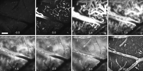
Fig. 3.
PIB kinetics in a PDAPP mouse model. Each frame is a maximum intensity projection of the fluorescence within a 3D volume of the brain of a live, transgenic PDAPP mouse, acquired with a Bio-Rad multiphoton microscope. Each 3D volume required 30 sec to obtain. Each frame is 615 × 615 μm wide and ≈150 μm deep. An i.v. injection of 10 mg/kg PIB was made just before t = 0 min. Note that the fluorescent compound appears within blood vessels almost immediately. At t = 0.5 min, the vessels appear larger in diameter and “fuzzy” as the compound crosses the BBB and enters the parenchyma. This is seen in the next time points, t = 1 and 1.5 min. Within 3 min, parenchymal plaques are labeled and remain labeled at t = 30 min (arrows). This transgenic animal had few and small parenchymal plaques and little CAA. The CAA was labeled within 0.5 min and remained labeled for >30 min. By 30 min, the diffuse fluorescence of the blood vessels and parenchyma was markedly reduced compared with the maximal brightness achieved at t = 1–3 min. Some nonspecific fluorescence remains, probably caused by the large dose of PIB given in this animal. (Scale bar: 100 μm.)

