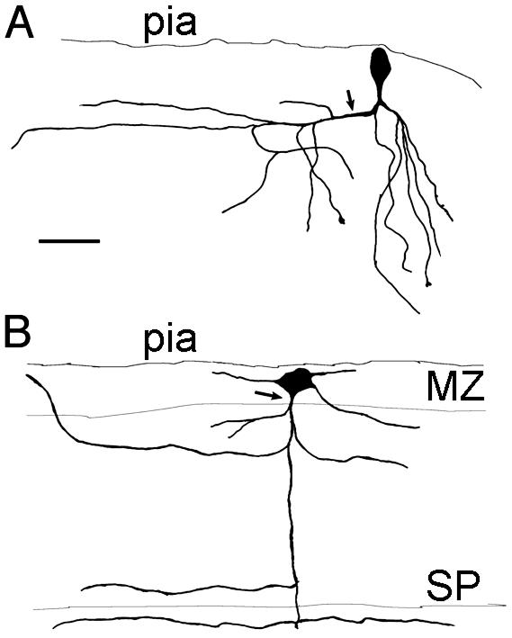
Fig. 1.
Morphology of pioneer neurons in the MZ labeled after CAV injections in the MGE and intracellular filling with Dextran-rhodamine. (A) A neuron with a fusiform, vertically oriented perikaryon. The axon is descending and gives rise to a collateral branch that extends toward the lateral side of the slice (arrow) and some descending branches. Dendrites are not shown. (B) A large multipolar neuron endowed with a descending axon that collateralizes in the CP and the SP. (Scale bar = 50 μm.)

