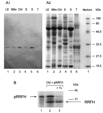
Figure 3.
Subcellular localization of RRFHCP. (A) Analysis of polypeptides from different spinach leaf subfractions. (A1) Western blot analysis of polypeptides (50 μg protein) from spinach leaf subfractions separated by SDS/PAGE with antibody (dilution of 1:1,500) against His6-partial RRFHCP. (A2) Coomassie blue staining of the corresponding gel. Lanes: 1, total leaves (LE); 2, mitochondria (Mito); 3, chloroplast (Chl); 4, chloroplast envelope membranes (E); 5, stroma (S); 6, thylakoid membranes (T); 7, molecular mass markers (low range; Bio-Rad). (B) Import of the [35S]methionine-labeled RRFHCP into isolated pea chloroplasts. The mixtures for the in vitro import reaction were analyzed by SDS/PAGE and fluorography. Lanes: 1, in vitro translation products of RRFHCP cDNA inserted into pCRII; 2, 35S-labeled RRFHCP (10-fold more than lane 1) was incubated with pea chloroplasts and chloroplasts were isolated after the import reaction; 3, the same as lane 2 except that the chloroplasts were treated further with thermolysin (TL) to remove proteins bound to the cytosolic face of the chloroplast outer envelope. The mature and precursor proteins are indicated as RRFH and pRRFH, respectively.

