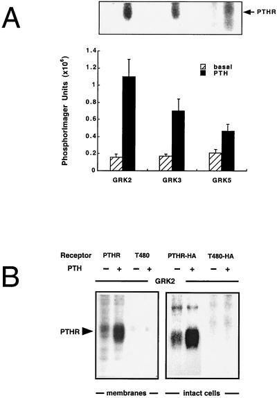
Figure 1.
Phosphorylation of the PTH receptor (PTHR) by GRKs in cell membrane preparations and in intact cells. Membrane preparations of Chinese hamster ovary cells stably expressing the PTH receptor (A) or COS-1 cells transiently expressing the wild-type or the C terminally truncated T480 PTH receptors (B, Left) were phosphorylated with 100 nM GRK2, GRK3, or GRK5 (A) or 100 nM GRK2 (B) for 30 min without or with 10 μM PTH. The reaction products were resolved on SDS-polyacrylamide gels, and autoradiograms of these gels are shown. 32P-incorporation was quantitated by PhosphorImager analysis (A). Phosphorylation by GRK2 was 1.1 ± 0.3 mol 32P/mol receptor (as determined by radioligand binding). Data in A represent the mean ± SE of four independent experiments. Phosphorylation of HA-tagged PTH receptors in intact [32P]orthophosphate-labeled COS-1 cells (B, Right) was achieved by incubating cells with 100 nM PTH for 5 min, followed by solubilization and immunoprecipitation of the receptors with 12CA5 antibody, SDS/PAGE, and autoradiography.

