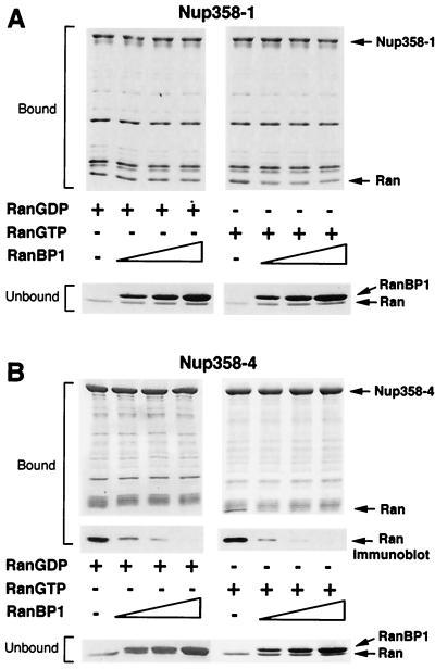
Figure 3.
RanBP1 competes with Nup-358–4 and, partially, with Nup-358–1 for Ran binding. (A) Glutathione-Sepharose beads with immobilized 0.25 μM Nup-358–1 were incubated with 0.8 μM RanGDP or RanGTP as indicated in the presence or absence of 1, 2, or 4 μM RanBP1. Bound and unbound fractions were visualized by SDS/PAGE and Coomassie blue staining. (B) Glutathione-Sepharose beads with immobilized 0.25 μM Nup-358–4 were incubated with 0.8 μM RanGDP or RanGTP as indicated in the presence or absence of 1, 2, or 4 μM RanBP1. Bound and unbound fractions were visualized by SDS/PAGE and Coomassie blue staining (Right) or Amido black staining after transfer to nitrocellulose membrane (Left). Ran also was visualized by immunoblotting with rabbit polyclonal Ran antibody (27) where indicated.

