Figure 5.
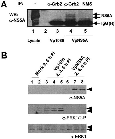
Physical and functional analysis of NS5A–Grb2 interaction in a virally infected system. (A) Anti-NS5A immunoblot comparing total lysate with anti-Grb2 or normal mouse serum (NMS) precipitated protein complexes from HeLa S3 cells infected with recombinant VV expressing NS5A (VpNS5A) or recombinant VV control (Vp1080) at 4 h postinfection. Lane 2 is an empty lane. The position of NS5A is indicated by the arrow. IgG(H) denotes the heavy chain of the mouse Ig. IP, antibody used for immunoprecipitation; WB, antibody for Western blot analysis. (B) ERK1/2 phosphorylation in HeLa S3 cells infected by recombinant VV expressing NS5A (VpNS5A) or recombinant VV control (Vp1080) at different time points postinfection. Cell lysates were fractionated with SDS/PAGE and then subjected to immunoblotting by using anti-NS5A [α-NS5A (A)], an antibody specific to the dually phosphorylated (Thr-202/Tyr-204) activated form of ERK1/2 [α-ERK1/2-P (B)] or anti-ERK1 [α-ERK1 (C)]. Position of NS5A and ERK1/2 is indicated by arrow.

