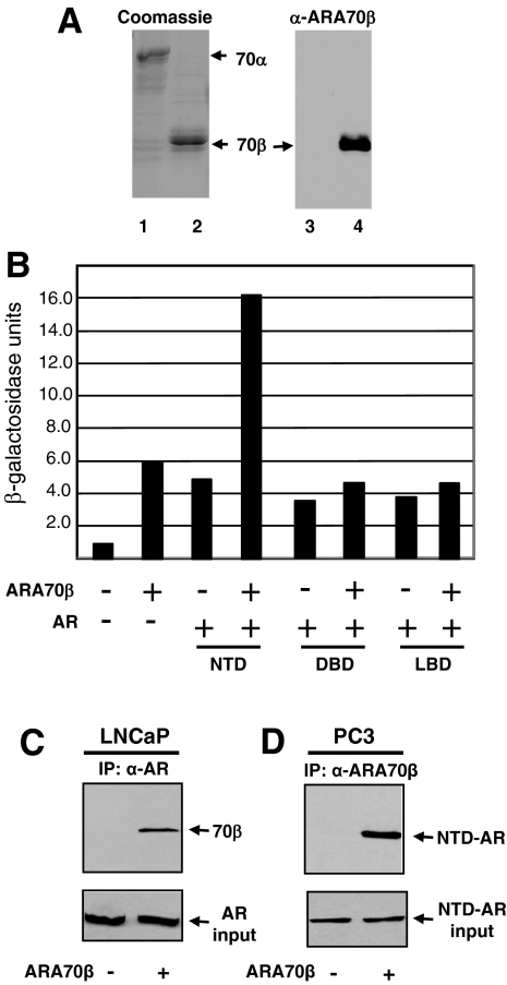
Figure 1.
ARA70β isoform-specific antibody production and interaction between AR and ARA70β. A: Coomassie blue-stained SDS-PAGE gel showing recombinant purified ARA70α (lane 1) and ARA70β (lane 2) (right). ARA70β isoform-specific antibody recognizes ARA70β (lane 4) and not ARA70α (lane 3) (left). B: Interaction of ARA70β with the AR NTD, AR DBD, and the AR LBD was analyzed using the yeast two-hybrid assay. The strength of interaction was determined by a quantitative liquid β-galactosidase assay after 24 hours of incubation in galactose media at 30°C. Note the threefold increase in β-galactosidase activity with the AR NTD and ARA70β compared to other domains of AR. C: ARA70β interacts with endogenous AR in LNCaP cell extracts by co-immunoprecipitation. LNCaP cells were stably transfected with pBabe-ARA70β or the pBabe (vector control) and whole cell extracts were prepared as described in Materials and Methods and incubated with an antibody against AR. Immune complexes were collected on protein A-Sepharose beads, washed, eluted, resolved by SDS-PAGE, and transferred to a membrane. The filter was probed with antibody against AR and ARA70β. D: ARA70β interacts with N terminus of AR in PC3 cells overexpressing ARA70β and transiently transfected with N terminus of AR, as determined by co-immunoprecipitation.

