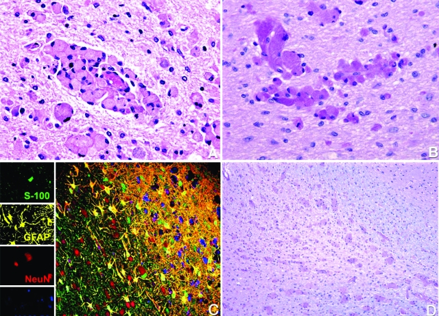
Figure 2.
Detection of globoid cells, gliosis, and demyelination in Krabbe-affected brain. A: Histological examination of the brain revealed massive accumulation of globoid cells in the white matter throughout the brain, principally around the microvasculature. Hematoxylin and eosin. Original magnification ×400. B: Globoid cells with fine granular periodic acid-Schiff-positive cytoplasm. Original magnification ×400. C: Multilabel confocal microscopy showing normal gray matter (left side) and extensive gliosis, loss of oligodendrocytes, demyelination, and numerous globoid cells in the white matter (right side). Images for individual channels (neurons with NeuN/Alexa 568, red; astrocytes with GFAP/CY3, yellow; oligodendrocytes with S-100/Alexa 488, green; and globoid cells with HAM56/Alexa 633, blue) are shown on the left with the larger merged image on the right. D: Extensive demyelination of the white matter. Luxol Fast blue. Original magnification ×100.

