Figure 5.
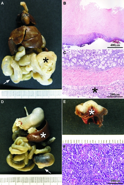
Causes of death. A: Macroscopic view of the thoracic and abdominal organs en bloc; there is a tumor at the cecum (black asterisk) whose growth has obstructed the intestinal traffic and dilated the small bowel (white arrow). B: Obstructive tumor showed areas of complete necrosis through the whole tumor thickness. C: Microscopic view of the large bowel from a case with intestinal obstruction; there are many inflammatory cells in the subserosal connective tissue (asterisk). D: Macroscopic view of the thoracic and abdominal organs en bloc; there is a white tumor (arrow) that has been bleeding; the spleen (white asterisk) is enlarged (compare with A) due to hematopoietic hyperplasia in response to chronic blood loss. E: On the cut surface, the white tumor showed a hemorrhagic surface. The intestinal lumen was filled with blood (white asterisk). F: The hematopoietic tissue of the spleen was hyperplasic in those cases with tumor bleeding. B, C, and F are H&E-stained sections.

