Figure 5.
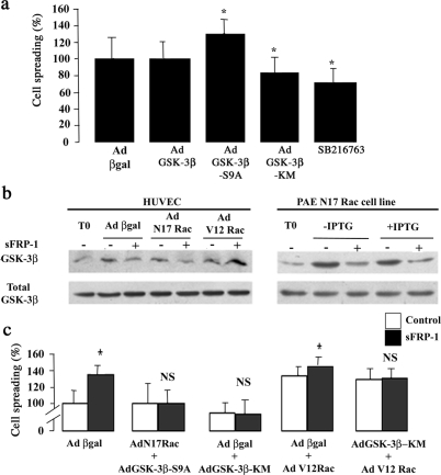
GSK-3β/Rac-1 pathway involved in sFRP-1-induced EC spreading. a: HUVECs were either infected with adenovirus expressing β-galactosidase, GSK-3β, GSK-3β-S9A, or GSK-3β-KM or treated with SB216763. After the treatment, a spreading assay was performed. Data are mean percent spreading ± SD. *P < 0.05 relative to control. b: HUVECs infected with adenovirus coding for β-galactosidase or N17Rac or V12Rac and PAE cell line expressing N17Rac under the control of an IPTG-inducible promoter were also treated or not with recombinant bovine sFRP-1 for 30 minutes in serum-free medium. After stimulation, the cellular proteins were analyzed by Western blotting with anti-phospho-GSK-3β (Ser 9) and total GSK-3β. The results shown are representative of three independent experiments. c: HUVECs were infected in a two-step process: first with adenovirus coding for β-galactosidase, N17Rac, or GSK-3β-KM, 6 hours before a second infection with adenovirus producing β-galactosidase, GSK-3β-S9A, GSK-3β-KM, or V12Rac. Eighteen hours later, cells were treated (▪) or not (□) with rb sFRP-1 for 30 minutes in serum-free medium. After stimulation, spreading assays on type I collagen were performed as described in the Materials and Methods section. Data are mean percent spreading ± SD. *P < 0.05 relative to control without recombinant bovine sFRP-1.

