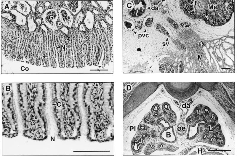
Figure 3.
Histological appearance of the mesonephric kidneys and lungs of African elephant fetuses. (A) Transverse section through the 115-day fetus showing the mesonephros with nephrostomes (N) opening into the coelom (Co) and connecting to the glomerulus (G) and associated collecting tubules (T). (B) High-power photomicrograph of one mesonephric nephrostome (N) clearly showing cilia (Ci). The cilia are characteristically angled inwards indicating the direction of flow of the filtrate. (C) Transverse section through the 139-day fetus showing mesonephros (M), metanephros (Me), dorsal aorta (da), and the posterior vena cava (pvc). The future spermatic vein (sv) leads directly from the mesonephros and the developing testis (not shown) to the posterior vena cava. (D) Transverse thoracic section through the 139-day fetus showing lung (L) and pleural cavity (Pl), heart (H) surrounded by pericardium, dorsal aorta (da), oesophagus (oe), and bronchus (B). [Bars = 0.25 mm (A), 0.16 mm (B), 0.64 mm (C), and 0.75 mm (D).]

