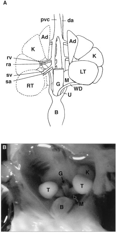
Figure 5.
Diagram and photograph of the ventral view of a dissected 166-day African elephant fetus. (A) This shows the short, straight spermatic vein (sv) and spermatic artery (sa). The right testis (RT) and Wolffian duct are displaced caudally (indicated by dotted outline) to show the course of the renal vein (rv) and the renal artery (ra) supplying the metanephric kidney (K) and the ureter (U). The right mesonephros has been omitted for clarity. The gut (G) has been removed, and the bladder (B) has been reflected caudally. Note that the adrenal glands (Ad) are large. The metanephric kidney is lobular as in the adult. The mesonephros (M) is regressing. (B) The large intra-abdominal testes (T) on the ventral aspect of the lobular metanephric kidney (K) are evident. The regressing mesonephros (M) is just visible. The gut (G) has been removed, and the bladder (B) has been reflected caudally.

