
Figure 2.

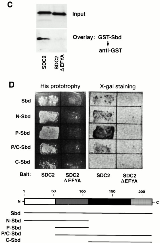
Characterization of the synbindin–syndecan-2 interaction by pull-down, coimmunoprecipitation, ligand overlay, and two-hybrid assays. (A) GST pull-down assay. Lysates from human 293 cells transfected with Myc-tagged syndecan-2 were pulled down with GST-synbindin fusion protein (a and b) or nonfused GST (c) and immunoblotted with anti–syndecan-2 mAb 6G12 (a) or anti-Myc polyclonal antibody A14 (b and c). In d, cell lysates were directly immunoblotted with anti–syndecan-2 mAb without pull-down. In each panel, three different lysates were tested: (lane 1) lysates from 293 cells transfected with a control vector; (lane 2) lysates from 293 cells transfected with Myc-tagged syndecan-2, which were undigested; and (lane 3) lysates from 293 cells transfected with Myc-tagged syndecan-2, which were digested with heparitinase. The arrowhead indicates the 34-kD syndecan-2 core protein. (B) Coimmunoprecipitation of synbindin and syndecan-2. The 293 cells were transfected with FLAG-syndecan-2 alone (lane 1), FLAG-syndecan-2 and Myc-synbindin (lane 2), or the FLAG-syndecan-2ΔEFYA deletion mutant and Myc-synbindin (lane 3). The cell lysates were immunoprecipitated with anti-Myc polyclonal antibody and immunoblotted with anti-FLAG antibody (c). The arrowhead indicates the syndecan-2 core protein. Note that the intact syndecan-2 was coimmunoprecipitated with synbindin (c, lane 2), whereas the syndecan-2ΔEFYA deletion mutant was not (c, lane 3). In a and b, the lysates were directly immunoblotted with anti-Myc antibody (a) and anti-FLAG antibody (b), respectively, to show that similar amounts of proteins were expressed. (C) GST-synbindin overlay assay. His-tagged recombinant proteins of intact syndecan-2 cytoplasmic domain (SDC2) and ΔEFYA cytoplasmic domain (SDC2ΔEFYA, right lane) were resolved on a 10–20% tricine gel, blotted, and overlaid with GST-synbindin (bottom panel) as described in Materials and Methods. (top panel) Ponceau S staining of the blot. (D) Two-hybrid assays to analyze the syndecan-2–binding site in synbindin. Four synbindin fragments (shown in the bottom panel) were tested with two syndecan-2 baits: one representing the intact syndecan-2 cytoplasmic domain (SDC2) and the other representing the ΔEFYA cytoplasmic domain (SDC2ΔEFYA). The interactions were scored by His prototrophy (left) and β-galactosidase activity (right).