Figure 4.
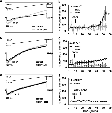
Electrophysiological recordings of membrane currents in HeLa-S3 cells before and after the application of cisplatin (CDDP). (a) Membrane currents of HeLa-S3 cells, elicited by a voltage ramp from −110 mV to +20 mV over 130 ms, before and with cisplatin (1 μM, n=5). (b) Time course of membrane currents (black line=measured at +20 mV; grey line=measured at −80 mV)) of HeLa-S3 cells using 1.8 mM calcium in the extracellular buffer. There was a clear increase in the currents at +20 mV after ∼40 min of application. After 55 min, membrane currents elicited at +20 mV were significantly different compared to the experiments where no calcium was added (compare panel d; *P<0.05; n=5). (c and d) Same procedure as in (a) and (b) but with no Ca2+ added to the external buffer. Note that after the application of CDDP, there was only a small increase in the current (n=5) over the time of the experiment at +20 mV and no change at −80 mV. (e and f) Same procedure as in (a) and (b) but using the KCa-channel blocker charybdotoxin (CTX; 3.3 nM). The examples shown illustrate that there is no change in the membrane current when the calcium-activated potassium channels are blocked, an indication that this is the main conductance activated by the entry of Ca2+.

