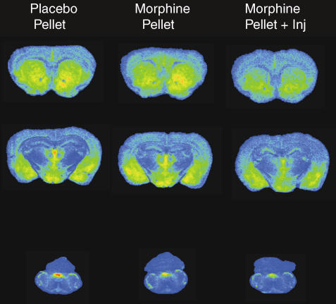
Figure 2.
DAMGO-stimulated [35S]GTPγS autoradiography in representative brain sections from mice implanted with placebo pellet, morphine pellet or morphine pellet+supplemental morphine injection. Sections were incubated with 0.04 nM [35S]GTPγS, 2 mM GDP and 10 μM DAMGO as described in Materials and methods. Areas examined densitometrically include: caudate-putamen, nucleus accumbens, cingulate cortex (row 1); thalamus, amygdala, hypothalamus (row 2); and NTS (row 3).

