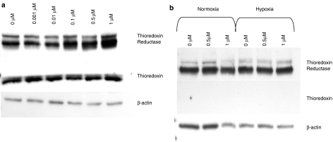
Figure 2.
Western blot analysis of Trx1 and TrxR1 levels. (a) HT29 cells and (b) MRCV cells were treated with varying concentrations of PMX464 for 72 h (final 48 h either under normoxic or hypoxic [1% O2] conditions; hypoxic data not shown for HT29). Blots were probed simultaneously for TrxR1 (∼58 kDa) and Trx1 (∼12 kDa), and stripped and reprobed for β-actin (∼42 kDa) as a loading control. PMX464, 4-(benzothiazol-2-yl)-4-hydroxycyclohexa-2,5-dienone; Trx1, thioredoxin; TrxR1, thioredoxin reductase.

