Figure 1.
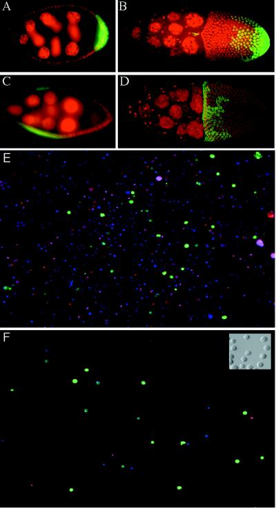
Marking and purification of follicle cell subgroups. (A–D) GAL4 expression patterns in egg chambers. Posterior is to the right. Nuclei are stained with propidium iodide, shown as red. Green fluorescence reflects GAL4-activated transcription of GFP. (A and B) A62 GAL4/UAS-GFP. (A) Stage 7 egg chamber showing GFP expression in posterior follicle cells. (B) Stage 10 egg chamber showing GFP expression in posterior follicle cells and in border cells (small anterior group). (C and D) 55B GAL4/UAS-GFP. Expression is seen in lateral follicle cells in stage 8 (C) through stage 10 (D) egg chambers. (E and F) Fluorescence images of cells released from A62 GAL4/UAS-GFP ovaries, before (E) and after (F) purification of GFP-positive cells by FACS. All nuclei stain blue with Hoechst 33342; the nuclei of dead cells stain red with propidium iodide (cells were restained after sorting). GFP fluorescence is shown in green. (Inset) Nomarski image of A62− cells after sorting.

