
Fig. 4.
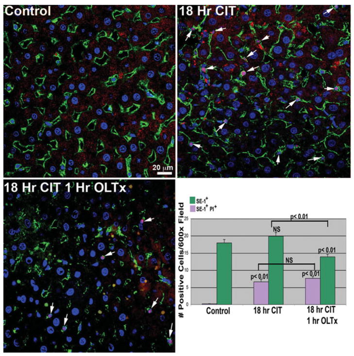
LSEC death in 18-hour CIT-stored and 1-hour post-OLTx livers. Livers were processed as described in Materials and Methods, and PI uptake by dead LSECs was evaluated by colocalizing PI nuclear signal (red, arrows) with SE-1+ cells in the sinusoid (green plasma membrane labeling). No PI labeling is observed in control livers. However, large increases in the pink-stained (red PI stain merged with blue nuclear Hoechst stain) LSECs is observed in both 18-hour CIT livers as well as 1-hour post-OLTx livers. Total LSEC (SE-1+) and PI+ LSEC numbers are indicated in the lower right-hand panel. As observed in our initial experiments (Fig. 1), numbers of SE-1+ cells are significantly decreased 1 hour post-OLTx. There is an increase in the percentage of SE-1+ cells also positive for PI uptake. Quantitation is the average of 3 animals from each condition; 12 separate 600× images from each animal were analyzed.