
Fig. 8.
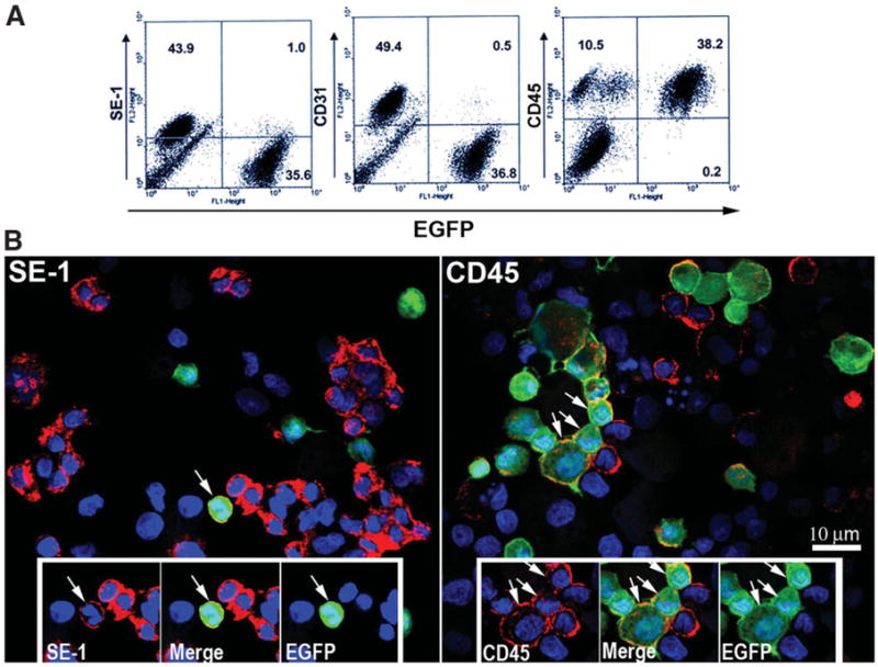
Isolation and characterization of origin of sinusoidal cells from an 18-hour CIT WT liver transplanted into an EGFP+ recipient. (A) Cells from a 3-day WT to EGFP+ recipient, 18-hour CIT, post-OLTX liver isolated by cationic magnetite were subjected to FACS analysis for surface markers SE-1 (LSECs), CD31 (large vessel endothelial cells and LSECs), and CD45 (leukocytes). In this particular animal, 1% of the total SE-1+ cells were EGFP+, 0.5% of the CD31+ cells were EGFP+, and 38.2% of the total CD45+ cells were EGFP+, confirming the general trend in the tissue shown in Fig. 6. (B) Cells subjected to FACS analysis were cytospun onto slides, counterstained with Hoechst nuclear stain, and evaluated using confocal fluorescence microscopy. As observed in the FACS and in tissue sections shown in Fig. 7, very few cells colocalize with both SE-1 and EGFP signal (arrow, left panel). Alternatively, many cells were both CD45+ and EGFP+ (arrows, right panel). Insets highlight positive and negative EGFP+ cells for each signal combination.