
Fig. 2.
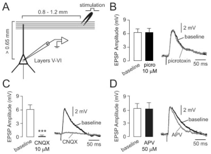
Electrical stimulation of superficial layers typically elicits a glutamatergic EPSP in deep-layer pyramidal neurons of the medial PFC. (A) Diagram illustrating the spatial arrangement of stimulating electrodes (layers I–II) and recording sites (layers V–VI). (B) Bath application of the GABA-A antagonist picrotoxin (10 μM) failed to change the amplitude of the evoked responses in all cells tested (n = 6). Left: bar graph summarizing EPSP amplitudes; right: example of the evoked response recorded before (black line) and after 5 min. of picrotoxin (gray line). Traces in this and subsequent figures are representative examples and not averages. (C) Bar graph (left) summarizing the effect of bath application of the AMPA/kainate antagonist CNQX (10 μM). The amplitude of the evoked response was gradually reduced and completely eliminated after 5 min. of CNQX in all cells tested (n = 8, ***P < 0.0001, paired t-test). Representative traces (right) showing the evoked EPSP before (baseline) and after 5 min. of CNQX. (D) Bath application of the NMDA antagonist APV (50 μM) failed to change EPSP amplitude in all cells tested (n = 7; left). Traces (right) recorded before (baseline) and after 5 min. of drug application illustrate the slight reduction of EPSP decay observed with APV.