
Figure 2. Co-localizations of PRL with rab11, rab7, and γ-adaptin in control and AdPRL transduced cells.
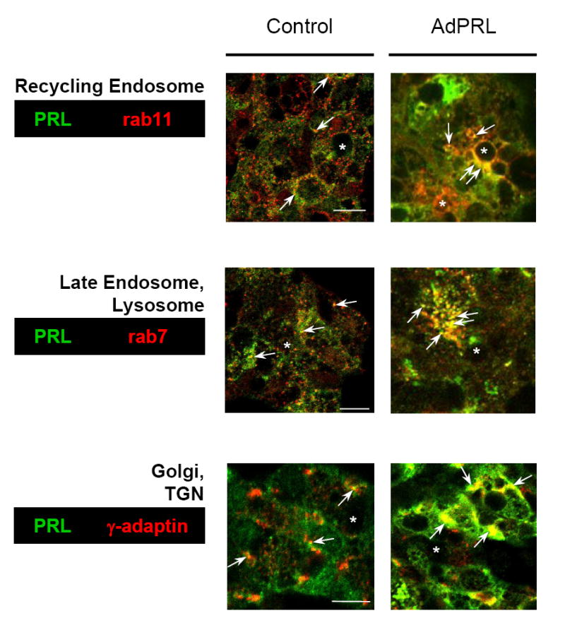
Control and AdPRL-transduced acini grown on Matrigel® coated coverslips were fixed and permeabilized with ethanol at −20°C (for rab7 and rab 11) or were fixed with 4% paraformaldehyde followed by permeabilization with 0.25% Triton X-100 (for γ-adaptin) then stained for PRL with guinea pig anti-prolactin antibody and rabbit anti-rab 7 or rab 11 or mouse anti-γ-adaptin antibody followed by FITC-conjugated donkey anti-guinea pig secondary antibody and rhodamine-conjugated goat anti-rabbit or mouse secondary antibody. In order to clearly visualize the co-localizations of PRL with rab11 and rab7 in AdPRL-transduced acini, unconjugated donkey anti-guinea pig IgG antibody was added to the FITC-conjugated donkey anti-guinea pig IgG secondary antibody at a 5: 1 ratio (*, apical/luminal regions; bar, 10 μm.)