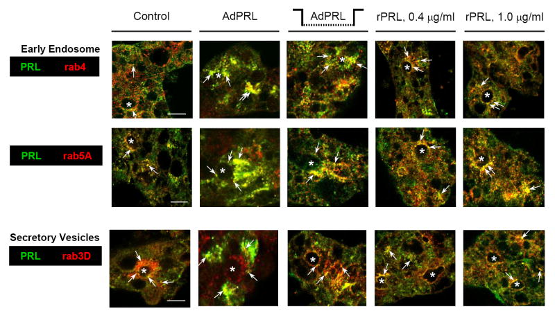
Figure 3. Co-localizations of PRL with rab4, rab5A, and rab3D.
Non-transduced acini grown on Matrigel® coated coverslips were cultured in the absence of added PRL; in the presence of either 0.4 μg or 1.0 μg recombinant rabbit prolactin; or in the presence of microporous culture inserts containing AdPRL-transduced acini. Additional acini were on Matrigel® coated coverslips were transduced with AdPRL. After rinsing, acini were fixed and permeabilized with ethanol at −20°C, then labeled with guinea pig anti-prolactin antibody and with rabbit anti-rab 4, anti-rab 5A, or anti-rab3D antibody. Secondary antibodies were FITC-conjugated donkey anti-guinea pig IgG and rhodamine-conjugated goat anti-rabbit IgG. For dual staining of AdPRL-transduced acini, unconjugated plain donkey anti-guinea pig antibody was added to the FITC-conjugated donkey anti-guinea pig IgG antibody at a 5: 1 ratio. (*, apical/luminal regions; bar, 10 μm.)

