
Figure 4. Secretion of internalized PRL by non-transduced cells.
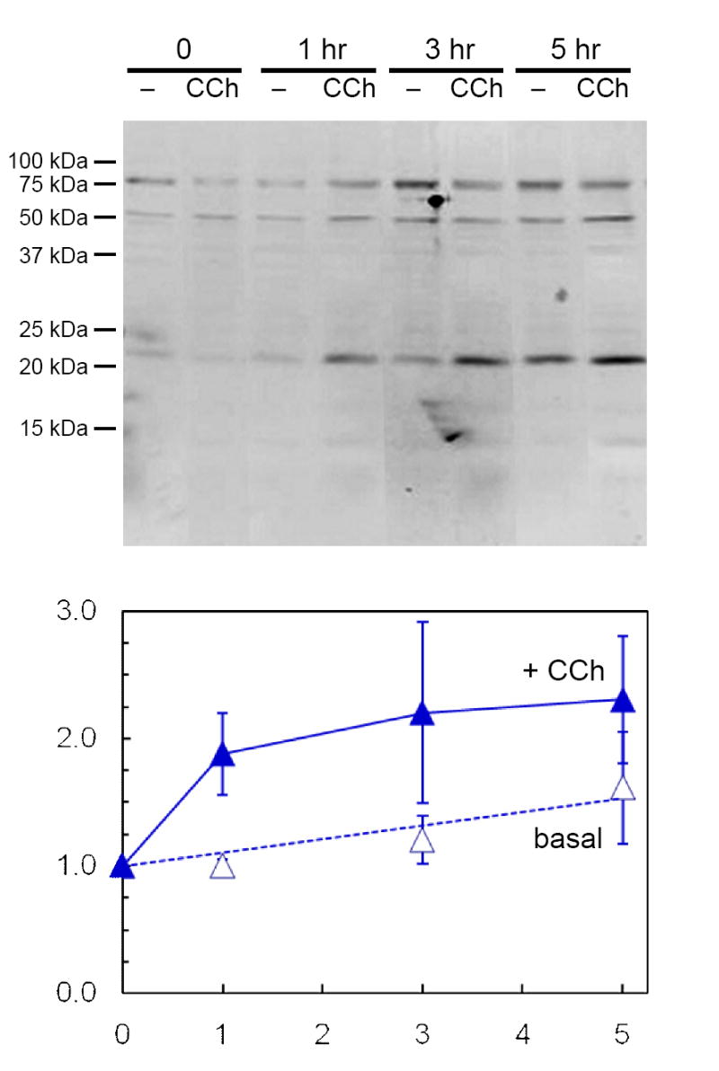
Non-transduced acini on Matrigel® coated 12-well plates (2×10⁶ cells/well) were incubated for 18 hr in the presence of microporous culture well inserts containing AdPRL-transduced acini. Following removal of the inserts and washing with PBS, fresh culture media with or without 100 μM CCh were added. After additional incubations for 1, 3 or 5 hr, the media were collected, centrifuged to remove debris, and concentrated 10-fold for analysis by Western blotting. Blots were probed with anti-prolactin antibody and IRDye 800-conjugated anti-guinea pig IgG antibody, then scanned and quantified as described in Methods. Amounts of 23 kDa PRL released are expressed relative to the 0 time value in the absence of CCh and represent the means ± sem for three separate preparations.