
Figure 6. EM-gold microscopy.
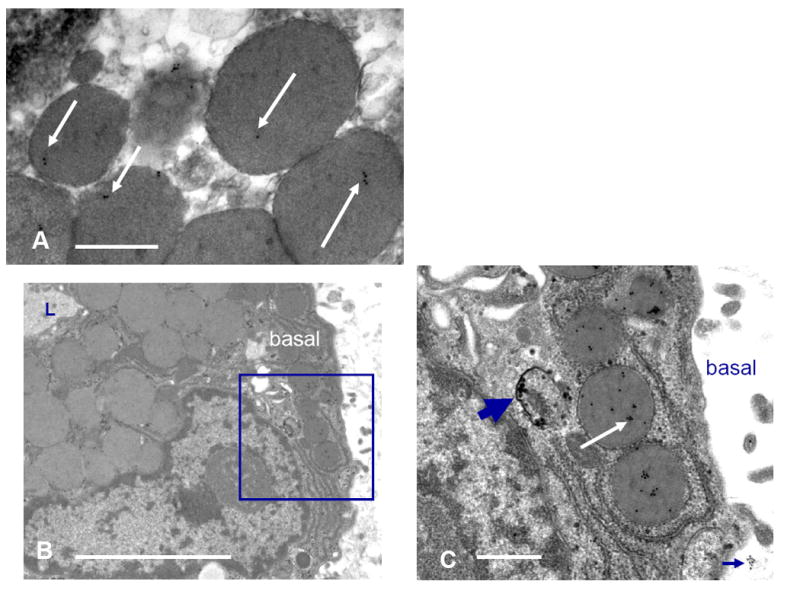
A. Control raft. Gold particles overlie secretory vesicles near the apical surface of acinar cells. B. and C. AdPRL-transduced raft. Micrograph B shows an acinar cell raft at low magnification with a portion of lumen (L) evident at upper left. Gold labeling is essentially absent from the apical secretory vesicles. The enclosed area is the basal cytoplasm of the acinar cell that is seen at higher magnification in micrograph C. The thick arrow indicates immunogold particles localizing PRL within a small vesicle, and the white arrow shows PRL localized within secretory vesicles. Occasionally small clusters of gold were evident just beyond the acinar cell basal surface (cluster at lower right), suggesting that PRL released at the basal surface associates with the subjacent extracellular matrix. (B Bar = 10 μ. C Bar =2 μ).