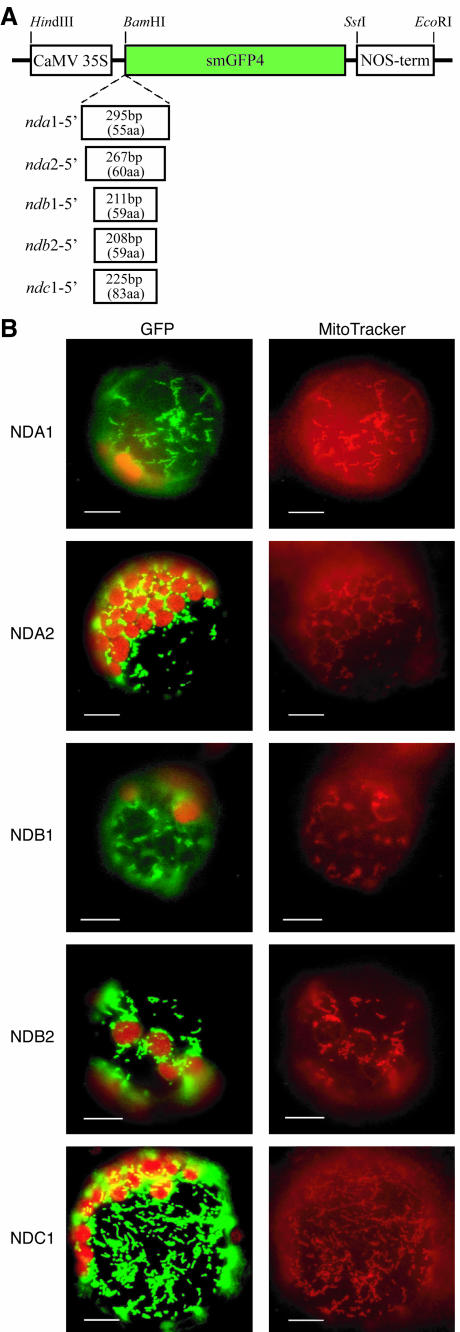
Figure 5.
Targeting analysis of gene products using GFP fusion proteins. The cDNA encoding the unconserved N-terminal part of each gene product, up to the start of the first nucleotide-binding motif, was fused to the reading frame of the smGFP4 reporter gene (A). The fusion proteins were expressed in transiently transformed protoplasts under the control of cauliflower mosaic virus 35S promoter and the NOS terminator. Location of the fusion proteins was analyzed by fluorescence microscopy (B). Images were taken using filter sets optimal for GFP and chlorophyll autofluorescence (left) and MitoTracker Red (right) and show for each construct the same protoplast. All GFP fusion proteins are observed in small cellular particles, which are also stained with the mitochondria-specific dye MitoTracker Red.

