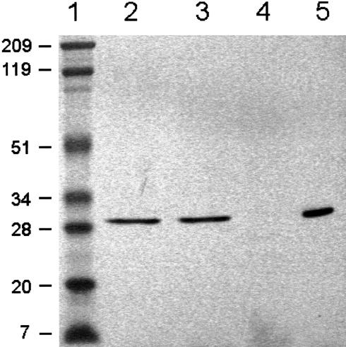
Figure 8.
Immunoblots showing localization of VuFeSOD in the soluble fraction of nodules (“cytosol”). Lane 1, Prestained molecular mass markers in kilodaltons. Lane 2, Cowpea nodule extract. Lane 3, Cowpea nodule cytosol fraction. Lane 4, Cowpea nodule membrane fraction. Lane 5, Purified recombinant VuFeSOD treated with thrombin. All lanes were loaded with 60 μg of protein, except lane 5, which was loaded with 0.2 μg of protein.

