
Figure 5.
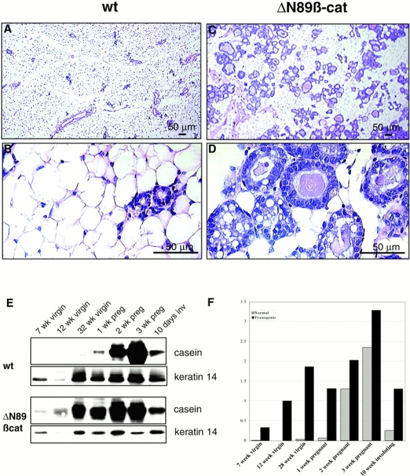
Histological and biochemical evaluation shows evidence of precocious epithelial differentiation in ΔN89β-catenin virgin mice. (A–D) Hematoxylin and eosin–stained sections of mammary gland from 6-mo-old virgin wt (A and B) and ΔN89β-catenin (C and D) littermates. Note the presence of vesicles in the transgenic samples. (E) Western blot of total proteins from the inguinal mammary glands at various stages of development, pregnancy (preg), and involution (inv) with anti–β-casein antibody and anti-keratin 14 antibody on the same blot, which serves as a control of epithelial content of the sample. (F) Graph of densitometry figures derived from the casein blots shown in E after normalization for keratin 14 levels. Note β-casein is expressed in ΔN89β-catenin but not wt virgin mice, and in all pregnant mice.