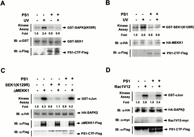
Figure 2.
PS1 inhibits UV-stimulated activity of SEK1 or MEKK1. (A) PS1 inhibits SEK1 activity. (B) PS1 inhibits MEKK1 activity. In A and B, HEK293 cells in 100-mm dishes were transfected with pcDNA3-PS1-Flag (4 μg), pEBG-SEK1 (1 μg), and pcDNA3-HA-MEKK1 (1 μg), as indicated. After 48 h of transfection, the cells were exposed to 80 J/m2 UV light, incubated further for 30 min, and lysed. For measuring GST–SEK1 activity, GST–SEK1 was isolated from the cell lysates using glutathione–agarose beads and then assayed for phosphorylation of GST–SAPKβ(K55R). For measuring HA-MEKK1 activity, the cell lysates were subjected to immunoprecipitation using anti-HA antibody. The immunopellets were assayed for MEKK1 activity by immune complex kinase assay. (C) PS1 does not affect ΔMEKK1-stimulated SAPK activity. HEK293 cells in 100-mm dishes were cotransfected with pcDNA3-PS1-Flag (4 μg), pcDNA3-SEK1(K129R) (1 μg), and pcDNA3-ΔMEKK1-Flag (1 μg) along with pcDNA3-HA-SAPKβ (1 μg), as indicated. (D) PS1 does not change Rac1-stimulated SAPK activity. HEK293 cells in 100-mm dishes were cotransfected with pcDNA3-PS1-Flag (4 μg) and pcDNA3-Rac1V12 (1 μg) along with pcDNA3-HA-SAPKβ (1 μg). In C and D, the transfected cells were lysed after 48 h of transfection. SAPK activity in the cell lysates was measured by immune complex kinase assay using mouse anti-HA antibody. IB, immunoblot analysis of transfected cells with the indicated antibodies.

