
Figure 5.
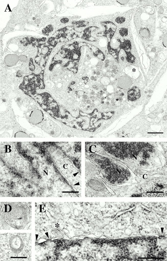
Ultrastructural analysis of a HeLa cell expressing B1Δrod by thin section EM. (A) Low magnification view of a whole nucleus. Bar, 1 μm. *Concentric membrane structure. (B–E) High magnification views. Bars, 300 nm. (B and C) A double membrane encloses the dense chromatin regions within lobules. Cytoplasm (C) and nucleus (N) are indicated. Arrowheads delineate nuclear pores. (D) Views of two concentric membrane structures in the cytoplasm. (E) Arrowheads delineate nuclear pores; *concentric membrane contained within the NE.