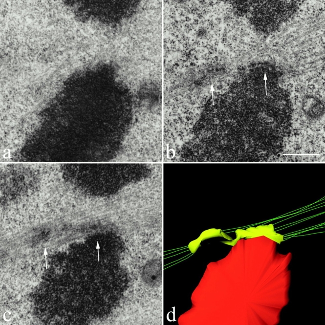
Figure 4.

Electron micrographs showing sequential-thick sections (a–c) through the merotelically oriented kinetochore of a lagging chromosome in a cell fixed at late anaphase. In panels b and c, kinetochore microtubules can be seen extending from the left side (left arrow) of the kinetochore toward the left spindle pole and from the right side of the kinetochore (right arrow) to the right spindle pole. (d) 3-D structure of the organization of kinetochore microtubules attached to the merotelically oriented kinetochore shown in panels b and c. The reconstruction was obtained from stereopairs for the two consecutive sections containing the kinetochore. The kinetochore is color encoded in yellow, the microtubule axes are green, and the chromatin proximal to the kinetochore is red. The figure clearly shows 5 kinetochore microtubules extending toward one pole and 11 kinetochore microtubules extending toward the opposite pole. It also clearly shows that the kinetochore is stretched laterally beyond the centromere region of the chromosome. For clarity, interpolar microtubules that pass near the kinetochore are not shown. A rotatable, 3-D file of panel d is available at http://www.jcb.org/cgi/content/full/153/3/517/DC1. Bar, 0.5 μm.
