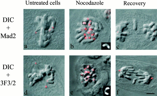
Figure 7.
Mad2 (a–c) and 3F3/2 (d–f) immunostaining in untreated, nocodazole-treated cells and cells fixed after 1 h recovery from a nocodazole-induced mitotic arrest. Overlays of differential interference contrast (DIC) and fluorescence images are shown. Mad2 and 3F3/2 staining are present on the kinetochores of unattached and unaligned chromosomes in prometaphase (a and d), are enhanced on the kinetochores of nocodazole-arrested cells (b and e), but are not present on the kinetochores of lagging chromosomes (or chromosomes normally moved to the spindle poles) in anaphase cells (c and f). The insets in the bottom right corners of panels b and e show higher magnifications of crescent-shaped kinetochores highlighted by a square in the pictures. Bar, 5 μm.

