
Figure 1.
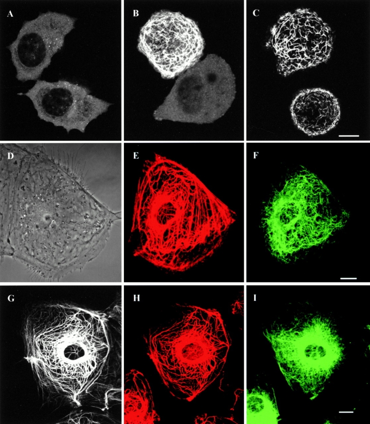
(A) No filamentous networks are seen in live SW13 vim− cells expressing GFP-K8 alone. Transfected cells display mainly diffuse and punctate fluorescence patterns. (B) Cotransfection with GFP-K8 and GFP-K18 frequently produces filamentous networks in live SW13 vim− cells. (C) When live SW13 vim− cells are observed after cotransfection with GFP-K8 and K18-myc, GFP-K8 coassembles with K18-myc to form squiggles and filamentous networks. (D–F) Phase–contrast image (D) of a live PtK2 cell expressing YFP-K8 (E) and CFP-vimentin (F). YFP-K8 displays a typical network of tonofibrils, which is distinct from the CFP-vimentin IF network. (G–H) GFP-K18 (G) also shows a typical tonofibril network in a live PtK2 cell. The same cell was processed for fixation and double immunofluorescence. Keratin is visualized with rabbit polyclonal anti–bovine tongue keratin and Alexa 568-labeled anti–rabbit IgG; and vimentin is visualized with mouse monoclonal anti–vimentin and Alexa 633-labeled anti–mouse IgG. GFP-K18 (G) is only incorporated into the endogenous network of tonofibrils (H) and not into the endogenous vimentin IF network (I). Bars, 10 μm.