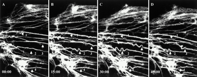
Figure 5.
Time-lapse observations of tonofibrils in live PtK2 cells transfected with GFP-K18. These fibrils exhibit extensive bending or wave-like movements. In many instances, these waveforms appear to be propagated along individual tonofibrils (arrows 2 and 3). In other cases, these waveforms disappear (arrows 1 and 4). Elapsed time (min:s) is indicated at the lower left of each confocal image. Video available at http://www.jcb.org/cgi/content/full/153/3/503/DC1. Bar, 5 μm.

