
Figure 2.
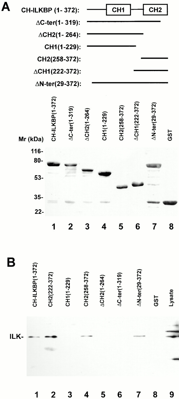
The CH2 domain mediates the interaction with ILK. (A) GST–CH-ILKBP fusion proteins were separated on SDS-PAGE (10 μg/lane) and detected by Coomassie blue R-250 staining. The numbers in parentheses indicate CH-ILKBP residues. (B) ILK binding. C2C12 cell lysates (100 μg) were incubated with equal amount (10 μg) of GST or GST fusion proteins containing CH-ILKBP sequences as indicated in the figure. GST and the GST fusion proteins were precipitated with glutathione-Sepharose 4B beads. ILK was detected by Western blotting with anti-ILK antibody 65.1. (Lane 9) C2C12 cell lysates (10 μg/lane).