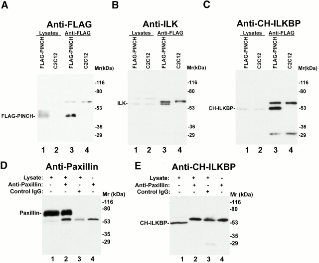
Figure 4.
Coimmunoprecipitation of CH-ILKBP and ILK with FLAG-PINCH. (A and B) FLAG-PINCH was immunoprecipitated from lysates of C2C12 cells expressing FLAG-PINCH. FLAG-PINCH immunoprecipitates (lane 3) or control precipitates (lane 4) were analyzed by Western blotting with anti-FLAG antibody M2 (A), anti-ILK antibody 65.1 (B), or anti–CH-ILKBP antibody 3B5 (C). Lanes 1 and 2 were loaded with lysates (7.5 μg protein/lane) of the FLAG-PINCH–expressing C2C12 cells and the control C2C12 cells, respectively. (D and E) Antipaxillin immunoprecipitates (lane 2) or control IgG precipitates (lane 3) were blotted with antipaxillin antibody (D) or anti–CH-ILKBP antibody 3B5 (E). (Lane 1) 10 μg lysates; (lane 4) 0.4 μg antipaxillin IgG.

