
Figure 6.
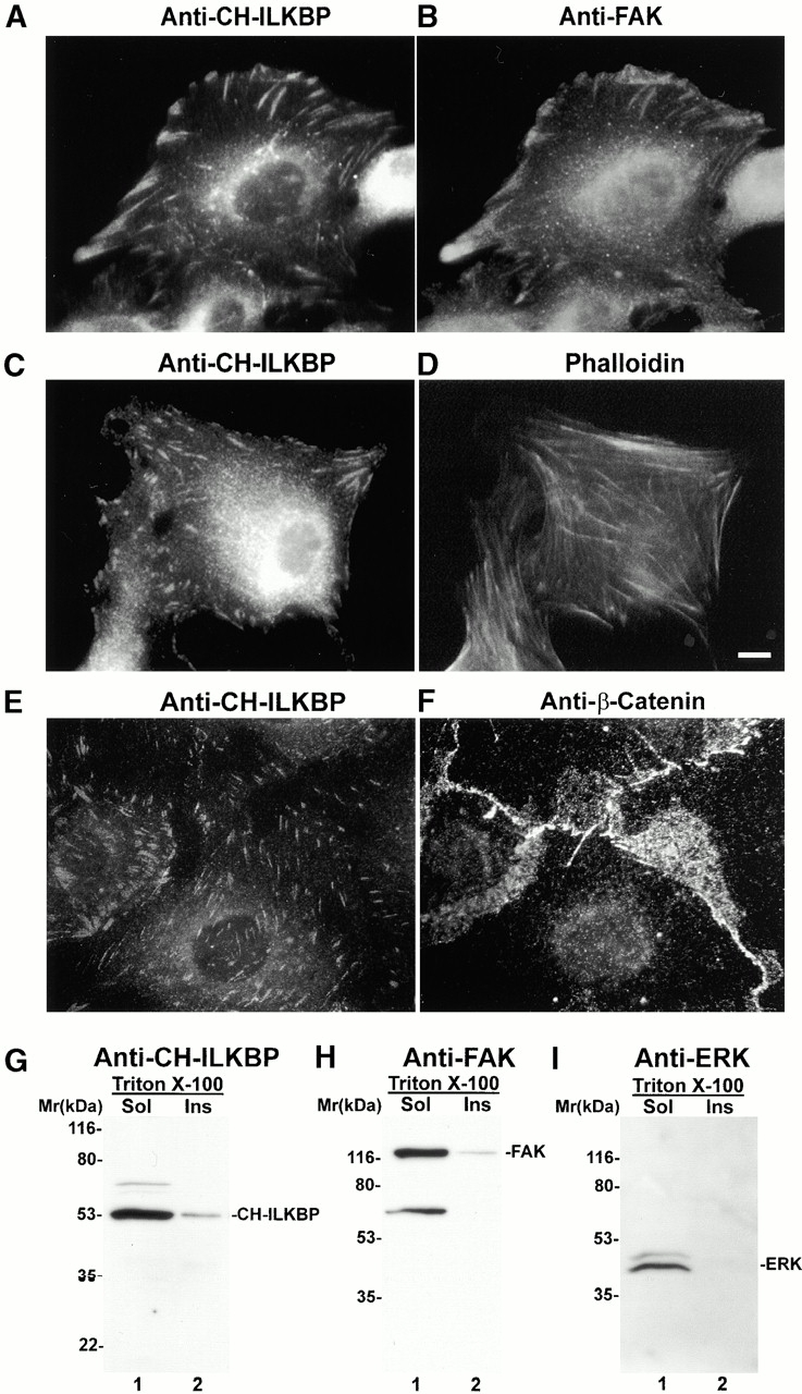
FA localization and cytoskeleton association of CH-ILKBP. (A–F) Subcellular localization of CH-ILKBP. Primary rat mesangial cells were stained with mouse anti–CH-ILKBP antibody 1D4 and rabbit anti-FAK antibodies (A and B) or anti–CH-ILKBP antibody 1D4 and phalloidin (C and D). We have also stained IEC18 rat intestinal epithelial cells, CHO cells, and mouse C2C12 cells with anti–CH-ILKBP antibodies and found that CH-ILKBP localizes to FA in these cells as well. Immunofluorescence images of IEC18 cells stained with mouse anti–CH-ILKBP antibody 1D4 (E) and rabbit anti–β-catenin antibody (F). (G–I) Association of CH-ILKBP with Triton X-100–insoluble cytoskeleton fractions. CH-ILKBP, FAK, and p44/42 ERK in the Triton X-100–soluble and –insoluble fractions were detected by Western blotting with anti–CH-ILKBP antibody 3B5 (G), anti-FAK antibody (H), and anti-p44/42 ERK antibody (New England Biolabs, Inc.) (I), respectively. Bar, 5 μm.