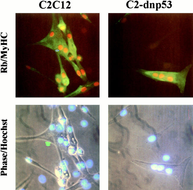
Figure 3.
pRb expression in differentiated myotubes. (Top) Indirect immunofluorescence performed on C2C12 and C2-dnp53 cells 72 h after the incubation in DM. Anti–pRB mAb was revealed by TRITC-conjugated antiserum and anti–MyHC serum by FITC-conjugated antiserum. (Bottom) Hoechst-stained nuclei pictures superimposed on the relative phase contrast images of the same fields shown above.

