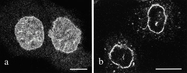
Figure 8.
(a). A confocal image of live PAM daughter cells in early G1 (20 min after telophase) showing the expression pattern of the GFP–COOH-terminal fragment of lamin B1 that lacks the central rod assembly domain. The mutant lamin is relatively uniform in its distribution throughout the nucleoplasm. (b) In contrast, GFP–wild-type lamin B is mainly located in the lamina region of the daughter cell nuclei at ∼20 min after telophase. Bar, 10 μm.

